The past few weeks, we have been learning about the cardiovascular system, a.k.a, the heart! We have looked at multiple diagrams learning the terms of the heart like the right and left atriums, pulmonary valves, and more!
We had a choice to dissect a cow, pig, or sheep heart. My group decided to dissect the pig heart.
After dissecting the heart we started to identify certain parts of the heart and state differences between the three species. One of the major differences between the three species is the size of the heart. In a cow, the heart is the largest because it needs to pump more blood throughout the cow's body. The pig also has a good sized heart because of the bigger body. The sheep has the smallest heart of the three species because with the smaller frame, it doesn't need to carry as much blood.
To further our knowledge in the heart and how it works, we checked blood pressure, examined each others heart rates with stethoscopes and EKG systems. After the discussion, we learned a lot from how heart rate fluctuates with what your body is like. If you are active and athletic, you are more likely to have a faster heart rate and if you are inactive, heart rate isn't as fast.
While learning about the heart, we also had to label a diagram of the heart consisting of the left and right atriums, left and right ventricles, vena cava, pulmonary veins, and more! This quiz wasn't as easy as I thought it would be and I didn't do that well. However, looking back now if I was to take the quiz again I think I'd be able to do better because my knowledge has expanded tons on the subject!
Anatomy & Physiology (:
Friday, March 30, 2012
Thursday, February 16, 2012
Dissection Time!
A few weeks ago we were to select groups and dissect the brain of a sheep. My group consisted of Gena, Zerek, and I. At first we were all pretty disgusted with the topic at hand. Zerek didn't want to touch it, let alone dissect it. After we got the necessary tools we had a choose of how to dissect the brain itself. The three ways were: sagittal, coronal, or transverse.
Sagittal- To cut into two separate pieces.(left and right)
Coronal- To cut in a vertical direction leaving you with a front and back piece of the brain.
Transverse- To cut horizontally which leaves you with a top and bottom
We chose to use the coronal cut to dissect the brain. However, before dissecting the brain itself, we had to remove the almost clear skin covering the top of the brain. This film is known as the dura mater. It protects the brain from getting damaged. After we got it dissected, Mr. Ludwig came and explained to us some of the parts of the brain, but unfortunately we ran out of time to discuss any thing further.
Here are some pictures of us dissecting the brain (:

Here are some pictures of us dissecting the brain (:

Tuesday, February 7, 2012
Memory!
From an early age, our memory begins forming and putting good and memories into our brain through the hippocampus. I expand more about what I learned in the glog I created. The knowledge I put together was very simple, however memory is very complex and has a lot of information to put all in one.
Wednesday, December 21, 2011
Bones, Bones, Bones!
For the section of skeletal system we had to split into groups of about 4-5 people. After that, we had to decide the section of the body we would like to memorize. My group consisted of Carla, Gena, Bri, Cassidy, and myself. Carla chose the midsection of the body such as the ribs and pelvis. Gena had the inside of the skull. Bri had the arms and fingers. I chose the bones on the outside of the skull, thinking it would be easy. After getting our assigned parts, I began looking at diagrams portraying certain parts of the skull and specific bones. After looking at multiple diagrams, I realized the skull has a ton of different bones! After about a day or two I started to get the hang of it and remember what certain bones were where. The bones I had to memorize are displayed in this picture!
Out of all these bones, I only missed about four.
Overall, I learned that there are multiple different bones not only in the body, but in the skull alone. I also learned not to wait last minute for memorizing certain bones! :)
Out of all these bones, I only missed about four.
Overall, I learned that there are multiple different bones not only in the body, but in the skull alone. I also learned not to wait last minute for memorizing certain bones! :)
Monday, December 19, 2011
Skeletal Muscle Fatigue-Eccentric Contractions and Muscle Damage
The muscle is stretched by outside forces or antagonist muscles frequently occur in normal conditions like walking down the stairs. When these contractions are repeated by someone out of shape, they make muscles weak and a characteristic muscle pain and tenderness; which become known a day or so after the workout. This type of damage to the muscle is linked with inflammation, hypercontracture of some fibers and protein loss from the damaged fibers. This type of pain and weakness can be the cause of some muscle symptoms in muscle diseases.
**Fun Fact**-Skeletal muscle fibers from rats with heart failure are more liable to mechanical damage, which shows that these muscles are more easily damaged during eccentric contractions.
Eccentric muscle damage includes characteristic changes to the sarcomeres with over- or under-stretched sarcomeres and wavy Z-lines. These are changes which have been explained by instability of sarcomeres in these situations. Evidence also shows that increased resting may act as a stimulus for inflammation and reduce Ca^2+ transients during these contractions, which add to the decreased force. A recent study has given researchers new information on possible early membrane damage after eccentric contractions. Vacuoles which were attached to the t-tubules were examined after contractions. Their figure could be held back by blocking the Na^+-K^+ pump. It was suggested that overstretched sarcomeres led to membrane tears, which allows the influx of ions like Ca^2+ and Na^+. The T-tubular-associated vacuoles are also a common thing in the damage of muscles and disease. Vacuoles and linked damage to t-tubules could hurt many cellular processes, such as, the action of exchangers and pumps in t-tubules, which can further hurt cellular dysfunction following the eccentric damage.
Overall, eccentric muscle damage can further damage other bodily processes if not taken care of in time. They are trying to find more and more things to cure this type of muscle damage.
Research Article:
http://www.medscape.com/viewarticle/444388_5
Tuesday, November 15, 2011
A&P Body Parts, Positions and More!
Just a few weeks ago we learned about the many different body positions and regions we have. I found out that our body can be classified with many different regions. My Popplet explains some of the regions and the anatomical position (: I also learned about the different body cavities that humans have. I further explain these in detail in a different popplet that I made. If you go to my site you can see the other popplet (:
Tuesday, November 8, 2011
The Integumentary System =]
The integumentary system is the skin. It has three primary functions which are to guard the body's physical and biochemical integrity, maintain a constant body temperature, and provide sensory information about the environment around them.
The skin is a big organ that is made up of all four tissue types. It is 22 square feet and 1-2 mm thick, and believe it or not, the skin is 10 pounds!
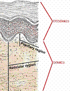
There are different parts to human skin starting with epidermis. The epidermis is the fake part of the skin and made of epithelial tissue. Within the epidermis there are four principle cells. The keratinocytes produce keratin, which helps keep the skin safe and protects underlying tissue from heat, microbes, and chemicals. It also has lamellar granules which gives off a waterproof sealant. The epidermis also includes melanocytes which make the brown pigment melanin. Langerhans' cells are also in the epidermis. These are epidermal macrophages that help start up the immune system. In the epidermis there are also Merkel cells, which function as touch receptors working with sensory nerve endings.
Layers of the Epidermis (:
Stratum Basal (Basal Layer) :
- Made of a single row of the youngest keratinocytes
- Deepest epidermal layer attached to the dermis
- The cells go through rapid division.
Stratum Spinosum (Prickly/Spiny Layer) :

- Cells are made of a weblike system of intermediate filaments that are connected to desmosomes.
- There are a lot of melanin granules and Langerhans' cells in this layer.
Stratum Granulosum (Granular Layer) :
- Very thin, 3-5 layers which big changes in keratinocyte appearance happens.
- Keratohyaline and lamellated granules gather in this layer of cells.
Stratum Lucidum (Clear Layer) :
- Very thin, see-through band that is superficial to the stratum granulosum
- Made of a few rows of flat, dead keratinocytes
- Only found in think skin.
Stratum Corneum (Horny Layer) :
- Keratinized cells
- Responsible for 3/4 of the epidermal thickness
- 3 different functions which include:
- Waterproof
- Protects against abrasions and penetration
- Helping the body closely insensitive to biological, chemical, and physical attacks.
What is the dermis?
The dermis is the 2nd major skin region which is made of strong, flexible connective tissue. The cell types include fibroblasts, macrophages, and sometimes mast cells and white blood cells.
Layers of the DERMIS =)
PAPILLARY LAYER :)
- The layer is made with areolar connective tissue with collagen and elastic fibers
- Its major surface contains peg like projections called dermal papillae
- Dermal papillae is made with capillary loops, Meissner's corpuscles, and free nerve endings!
RETICULAR LAYER (:
- This layer is responsible for about 80% of the skins thickness.
- The layers of collagen in this layer make the skin stronger and more resilient.
- The elastin fibers provide stretch-recoil properties.
Last but not least is the hypodermis. It is the subcutaneous layer which is deep to the skin. It is made of adipose and areolar connective tissue.
Resource!
Subscribe to:
Comments (Atom)


